Researchers at Imperial College London, UK have demonstrated for the first time that microscopic bubbles of gas can be manipulated using sound waves. The new “acoustical tweezers” overcome certain limitations of their optical cousins (such as not propagating well through opaque tissues), and could therefore enable a host of biomedical applications.
Microbubbles are already routinely employed in medicine as contrast agents in applications such as sonography. They could also be ideal in emerging ultrasound therapies such as tumour and kidney-stone destruction; stroke management; and delivering drugs to areas of the body that are hard to reach using conventional techniques. The first step, though, is to develop better ways of manipulating them. “It is important to be able to control the position of a microbubble in a contactless fashion in its native and complex environment to precisely analyse its response to ultrasound,” says Diego Baresch, the study’s lead author. “This is what we have now demonstrated in our work.”
Acoustic vs optical tweezers
Baresch began working on acoustic manipulation as a PhD student at the Université Pierre & Marie Curie in Paris. There, he and his colleagues showed that specially-structured sound waves could produce a trapping force on a solid object in all directions of space. “This implies that the object can be pulled in the direction opposite to the propagation of the sound waves,” he explains. “This is counter-intuitive for scientists because of momentum conservation rules and is exactly what Arthur Ashkin achieved in 1986 with lasers to develop the device known as ‘optical tweezers’, for which he received the Nobel Prize in Physics in 2018.”
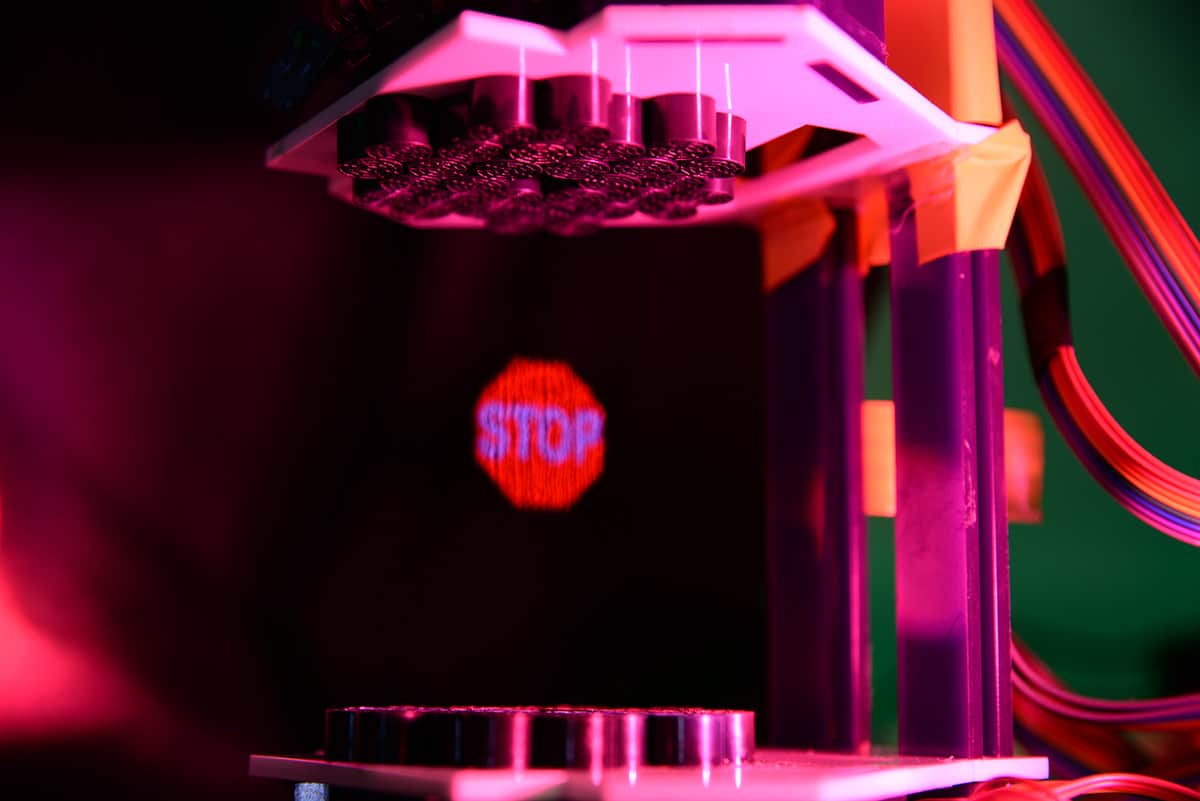
Whereas optical tweezers used light to trap and manipulate molecules, particles and cells, Baresch explains that the acoustic version employs a helicoidal or “vortex” beam of ultrasound. This, he says, has several benefits for medical applications. “We now know that the force exerted on objects at moderate acoustic pressures is orders of magnitude higher compared to optical forces,” he tells Physics World. “This radiation force could be used to probe a wealth of systems – for example, intercellular forces in biological cells, cellular adhesion forces and many other mechanisms involved in tissue development.”
Ultrasound can also penetrate deeper into opaque media such as biological tissue than optical waves, adds study co-author Valeria Garbin. This is an advantage over laser light, which is highly attenuated and can cause irreversible damage to cells – an effect that Ashkin termed “opticution”.
Single-beam acoustical trap
To test their technique, the researchers used a single-beam acoustical trap to manipulate microbubbles in three dimensions through three-centimetre-thick layers of elastic materials. They found that they could indeed move the microbubbles through this test material, which was designed to mimic biological tissue. They also showed that they could controllably release nanoparticles contained in the microbubbles using a second acoustical trigger, proving that their approach could be used to deliver therapeutic agents.

Making images with sound
The work opens the way for acoustical tweezers to be used in a broad range of applications in biology and medicine, say the researchers, who report their work in PNAS. “We hope we will now have the chance to employ this technique in collaboration with other experts in biophysics and biomedicine and take it a step further,” Baresch says.
