From proton-based cancer therapy to small-animal PET scanners, US technology company H3D is seeking out growth opportunities in the medical imaging market

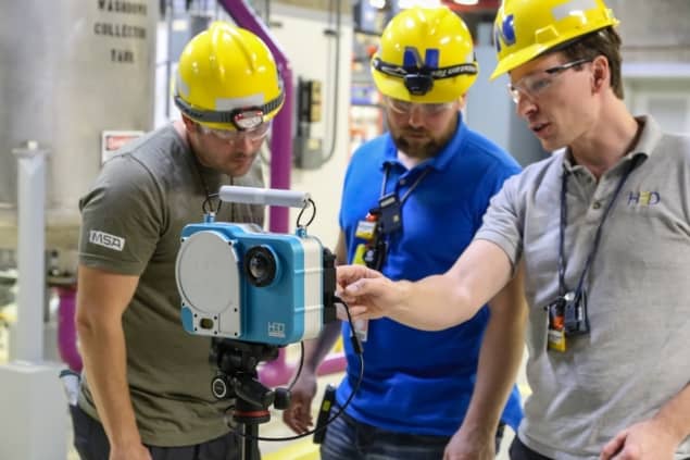
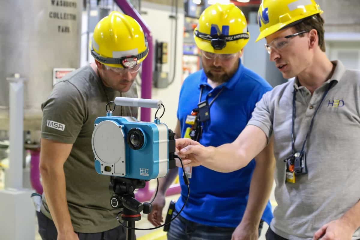
H3D is a US technology and research-focused business that provides high-performance imaging spectrometers for real-time identification and localization of gamma-ray sources. Over the past decade, the Ann Arbor, Michigan-based manufacturer has made its name serving a diverse base of end-users with specialist measurement solutions for gamma-ray imaging and spectroscopy. That customer base includes government agencies and nuclear first-responders, radiation safety officers at nuclear power plants, and international inspectors tasked with safeguarding and compliance in accordance with nuclear non-proliferation treaties.
“Our commercial instruments are based around cadmium zinc telluride (CZT) radiation detectors – a technology that combines industry-leading energy resolution and spatial resolution with room-temperature operation,” explains Willy Kaye, founder and CEO of H3D. As well as 10 full-time, PhD-level staff working on CZT device development and optimization, H3D draws on a broader pool of engineering and technical talent across the regional supply chain – most notably the Detroit car industry. “The emphasis at H3D is on robust, fully integrated radiation measurement solutions ready for field deployment,” Kaye adds. “To make this possible, we leverage expertise from a range of industry partners to ensure best practice in areas like manufacturability, packaging, mechanical testing, control electronics and software.”
The logic of diversification
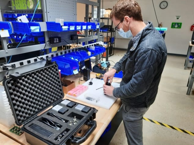
If that’s the back-story, the next chapter in H3D’s development is already taking shape, building on those solid foundations to address CZT growth opportunities in the medical imaging market. Front-and-centre in H3D’s diversification effort is the M400 Series, a compact and customizable module capable of high-resolution gamma spectroscopy in a range of medical imaging applications. “The M400 is an off-the-shelf commercial product that can be easily ‘tiled’ to form imaging arrays with enhanced sensitivity,” says Kaye. “We anticipate broad applicability across multiple imaging modalities and clinical use cases.”
Right now, H3D’s engagement with the medical imaging community manifests in a targeted programme of R&D collaboration. It looks like a win-win: proof-of-concept experimental projects allow H3D engineers to gather custom requirements at scale, while simultaneously educating medical imaging specialists about the versatility and capability of CZT technology. One such collaboration is with the Maryland Proton Treatment Center in Baltimore. This six-year R&D effort, funded by the US National Institutes of Health (NIH), is evaluating the potential of CZT imaging in proton cancer therapy, an advanced form of radiotherapy that allows precise radiation delivery to complex tumour volumes while sparing healthy tissue and organs-at-risk.
Specifically, the M Series forms the basis of a high-energy, high-flux spectrometer that’s able to image high-energy gamma rays produced during proton therapy (the spectrometer being tiled up with 16 M400 units – i.e. 64 CZT crystals and a total crystal volume of 310 cm3). Although this is still early-stage evaluation work, the long-term objective is an on-board imaging system to ensure that the bulk of the radiation payload from the proton treatment beam is deposited into the tumour rather than adjacent healthy tissue.

“The prompt gamma-ray emissions from proton interactions with tissue can be used to monitor the proton beam in vivo, providing real-time knowledge of tumour location, beam position, and beam penetration depth during treatment,” explains Hao Yang, a product development engineer at H3D.
“Worth noting,” he adds, “that the readout system is optimized for the high-flux conditions of proton cancer therapy, though ultimately it’s the system integration aspects that will be key to successful commercial engagement with the proton therapy OEMs.”
Functional imaging
In a separate collaboration, H3D engineers are investigating opportunities in small-animal positron emission tomography (PET), a functional imaging technique that uses radioactive tracers to track changes in the metabolism of diseased tissue (e.g. cancerous tumours) and physiological processes such as blood flow and chemical absorption. As such, small-animal PET provides a key imaging modality for the preclinical evaluation of new drugs, allowing biomedical scientists to track a drug’s behaviour over time versus a range of metrics such as treatment efficacy, biodistribution, toxicity and excretion. These small-animal studies, in turn, inform the regulatory approvals process ahead of advanced clinical trials in human patients.

With this in mind, H3D has developed a prototype imaging detector (based on four specially arranged M400 modules) for applications in small-animal PET studies. The custom system, which is currently being evaluated by researchers at the University of Illinois at Urbana-Champaign, is capable of achieving 0.5 mm spatial resolution (versus 1 mm for the best commercial scintillators). “This is a significant step forward for small-animal PET and the best positional resolution of any traditional PET detector,” claims Kaye. Separately, the Urbana-Champaign scientists have also purchased 50 M400 modules to evaluate novel detector arrays for next-generation functional imaging systems.
Another application of interest for H3D is the monitoring of radioisotope distribution within the body during diagnostic or therapeutic medical procedures. The gamma-emitting technetium-99m, for example, is used for functional imaging of the skeleton and a range of organs (including the heart, liver, kidney and gall bladder), while iodine-131 is widely deployed for the treatment of an overactive thyroid gland or as a post-surgical follow-up when treating thyroid cancer.
In this scenario, H3D’s gamma-ray imaging spectrometers enable clinicians to validate their organ transport models for radiopharmaceuticals by providing precision overlay of gamma and optical images of the patient in real-time. “While we are confident that we have developed a truly unique measurement tool,” concludes Kaye, “as sensor developers we will need to rely on the creativity of our potential partners to figure out how to improve patient outcomes based on this technology.”
Product focus: the M400 Series

H3D’s M400 Series is a custom integrable CZT detector module for high-resolution gamma spectroscopy and imaging. The product, which can be used as a single detector or tiled together for increased sensitivity, is suitable for a range of OEM system applications including drones, robots and medical imaging arrays. Technical specifications include:
- Spatial resolution: <0.5 mm (≥140 keV)
- Energy resolution: ≤1.1% FWHM at 662 keV (coincident interactions combined); enhanced resolution version also available (≤0.8% FWHM at 662 keV)
- Crystal volume: >19 cm3 CZT (484 detector pixels)
- Spectroscopy range: 50 keV to 3 MeV
- Compton imaging range: 250 keV to 3 MeV (optional)
- Dimensions: 10.2×5.3×5.3 cm and 0.6 kg
