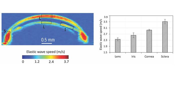
A new technique evaluates the biomechanical properties of the eye with much better elastic resolution than current methods, raising hopes for more effective diagnostics and therapies. The new approach, put forward by researchers from the University of Houston in Texas in the US, goes by the name of multifocal acoustic radiation force reverberant optical coherence elastography, and might also improve our understanding of how different eye components function.
Eyes are extremely complex organs made up of several types of specialized tissue that help maintain intraocular pressure and thus allow us to see clearly. This normal biomechanical function is disrupted, however, in diseases such as keratoconus and glaucoma. Understanding how these changes happen is vital for diagnosing and treating ocular pathologies, but for that to be possible, clinicians need to be able to assess biomechanical properties such as stiffness. Current methods of doing this have important limitations. Magnetic resonance imaging (MRI), for example, is costly and requires patients to remain still for long periods. This includes not moving their eyes, since even small movements can lead to errors in measurements.
Creating 2D or 3D elasticity maps
In the new study, researchers led by biomedical engineer Kirill Larin employed a technique called reverberant optical coherence elastography (RevOCE) to measure the elasticity or stiffness of eye structures with high resolution. RevOCE involves using a low-power light source to scan a specific volume of the eyeball and then detecting the complex interference patterns produced by vibrating mechanical waves that subsequently propagate through the eye tissue. These patterns are then used to create 2D or 3D elasticity maps of the region studied.
The drawback is that the complex interference patterns of mechanical waves are difficult to generate, and the usual methods of doing so require the source of the waves – a mechanical shaker – to contact eye tissues directly. This can be very uncomfortable for the patient.
Larin and colleagues’ innovation was to generate reverberant shear waves with RevOCE in a much less invasive way, without compromising on resolution. Their new system comprises a multifocal acoustic radiation (ARF) system with an ultrasound generator coupled to an array of acoustic lenses. These lenses focus the ultrasound waves to produce three distinct ARF beams spaced a few millimetres apart. These beams are then sent through a target area within the eye, where they induce vibrating shear waves that can be detected by an optical coherence tomography device and post-processed to reconstruct a 3D elastography map of the entire eyeball.
Comprehending overall eye function
The researchers tested their technique on ex vivo mouse eyeballs and confirmed that it could produce shear wave speed maps within different structures of the eye, including the cornea, iris, lens, sclera and retina. They found that the speeds of the waves were different in eye components such as the apical region of the cornea and the pupil of the iris. That is perhaps not surprising, but they also found that the wave speeds differed within different regions of the same component, such as the apex and periphery of the cornea. This implies that apparently uniform structures in the eye have non-uniform biomechanical properties – something that may be important for their healthy function.
“Our technique provides valuable insights into how different components of the eye maintain their relative stiffness,” Larin says, “and the insights we have obtained will aid in developing a detailed understanding of how these components interact with each other.” Indeed, he adds that the information garnered during this work, which is detailed in the Journal of Biomedical Optics, could help researchers and clinicians analyse the biomechanical relationships within individual ocular components and among different component. This will be important for understanding overall eye function in healthy eyes, as well as for diagnosing and monitoring pathologies.

Solitons from new fibre laser could improve eye surgery
“Many eye diseases and conditions, such as glaucoma, affect multiple ocular components simultaneously,” he tells Physics World. “By assessing the biomechanical properties of the whole eyeball, as in our approach, it becomes possible to detect these diseases in their early stages, even before symptoms manifest in specific components. Early detection can lead to more effective treatments and preservation of vision.”
What is more, monitoring how the biomechanical properties of the entire eyeball change over time is essential for tracking disease progression. “Some diseases may affect one component first before impacting others,” Larin says. “By evaluating the entire eyeball, clinicians can better assess disease evolution and make timely adjustments to treatment plans.”
The University of Houston team now plans to further refine and validate its modified RevOCE technique. This will involve conducting in vivo studies and exploring applications in monitoring disease progression and treatment outcomes in different animal models, and, eventually, in humans. “We are also interested in expanding its use to other areas of the body in addition to the eye – for example, for non-invasively evaluating the biomechanical properties of deep tissues,” Larin reveals.
