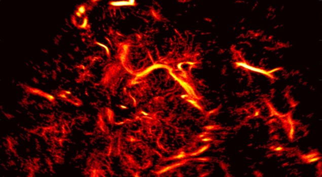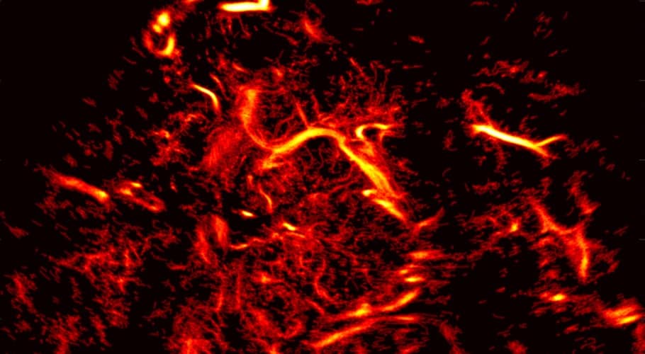
Researchers at the laboratory Physics for Medicine Paris have performed the first microscopic mapping of the vascular network in the human brain. The team used transcranial ultrafast ultrasound localization microscopy (ULM) of intravenously injected microbubbles to capture intracranial blood flow dynamics with a resolution of around 25 µm.
This breakthrough in ULM technology, described in Nature Biomedical Engineering, may help expand the fundamental understanding of brain haemodynamics and shed light on how vascular abnormalities in the brain are related to neurological diseases and disorders.
“By revealing both the human cerebral vascular anatomy and flow dynamics at microscopic scales, transcranial ULM is very likely to become of major importance for the management of cerebrovascular diseases in the future,” state the researchers. “Its low cost, ease of use, sensitivity and quantification capabilities, in combination with an improvement of almost two orders of magnitude in resolution compared with current clinical modalities, could transform the workflow of cerebrovascular patient management.”
Vascular dysfunctions are the underlying cause of many neurologic disorders, but diagnosing and treating these diseases has been difficult due to limitations of CT-angiography and MR-angiography, today’s most commonly used cerebrovascular imaging methods. These modalities are unable to detect capillaries with micron-sized diameters and do not provide dynamic information at the different spatial scales of the vascular network.
First-in-human study
Principal investigator and laboratory director Mickael Tanter and colleagues examined three patients receiving treatment at the University of Geneva’s clinical neurosciences centre, plus one healthy volunteer. The participants were intravenously injected with an ultrasound contrast agent consisting of a suspension of tiny microbubbles (SonoVue), which improves signal-to-noise ratio by enhancing the echogenicity of blood.
The researchers acquired transcranial images using an ultrafast programmable scanner and a phased array ultrasonic probe placed in front of the temporal bone window (the thinnest area of the lateral skull). For data acquisition, the ultrasound is on for 1 s, pulsing diverging waves at 4900 Hz from four virtual sources while acquiring data, then the ultrasound is off for 1 s. This process loops for a desired duration ranging from 24 s to 2 min 15 s. To process the signals, the team developed methods to measure and correct for skull-bone aberrations and motion artefacts.
The researchers employed a vesselness filter, an image processing filter optimized to improve the contrast of vascular structures, to make microbubbles moving in time appear as tubular structures in a 3D matrix. After building vascular maps, they performed flow quantification and estimated functional resolution, and used computer-generated animation to help visualize the functional information extracted from the bubble positions and tracking data.
By determining the positions of millions of microbubbles within a few seconds, the researchers were able to map the anatomy of the vascular network to functionally characterize blood-flow dynamics with microscopic resolution. They were also able to detect blood vortices in a small deep-seated aneurysm in a patient.
The team achieved this feat by combining several techniques. “The first technique is ultrafast imaging, which provides a tremendous amount of data in a very short time, and enables us to discriminate the acoustic signature of each individual microbubble,” lead author Charlie Demené explains in a press statement. “Then, ultrasound localization overrides the resolution limits inherent to wave physics: the image of a tiny object is a blurry spot which is larger than the actual object. But if this object is isolated, we can reasonably assume that its exact location is the centre of the blurry spot.”

Ultrafast 3D ultrasound wins journal citations prize
“In our case, the microbubbles circulating in the blood stream play the role of isolated objects and allow us to recover the exact location of each blood vessel,” he adds. “Finally, recording the echo of each microbubble gives access to the history of the wave coming from this micron-sized object, and hence enables us to recover what occurred during the propagation of the wave through the skull in order to correct its perturbations.”
“The next step will be to demonstrate the enhanced diagnostic capabilities of this new brain angiography technique for the survey of brain aneurysms and post-stroke recovery,” Tanter tells Physics World. “On the technical side, we are currently expanding this imaging to full 3D imaging. We are collaborating with Iconeus, a spinoff company of our lab, to develop a commercial product using transcranial ULM technology.”
