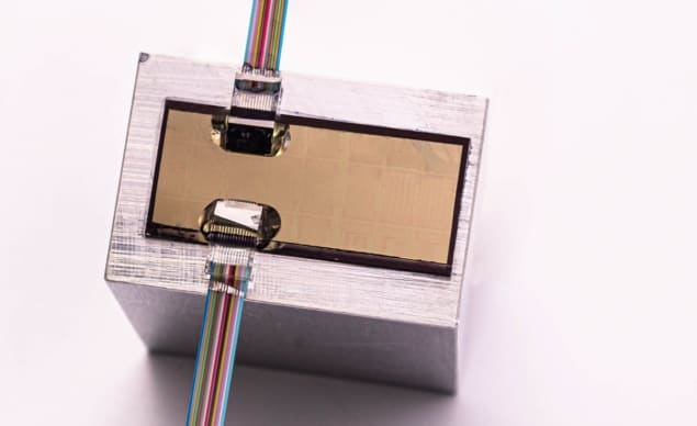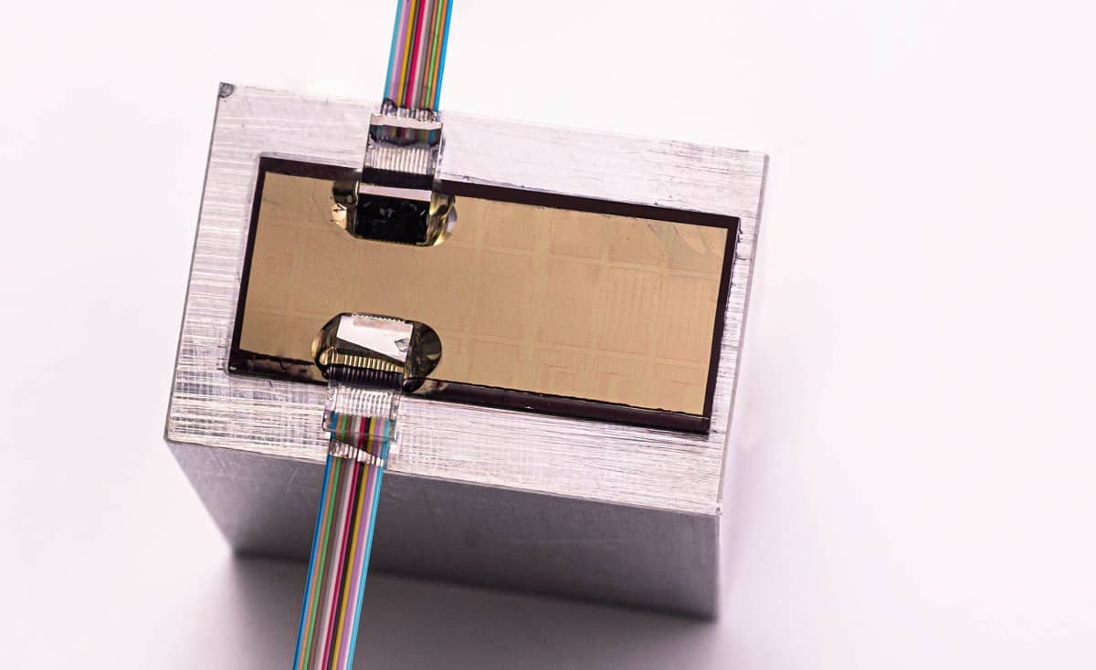
A highly sensitive optomechanical ultrasound detector integrated onto a silicon photonic chip has been developed by researchers in Belgium and Germany. The team, led by Wouter Westerveld at the Interuniversity Microelectronics Centre (IMEC) in Leuven, showed that its device is 100 times more sensitive than state-of-the-art piezoelectric detectors of identical sizes. Their design could substantially improve the performance of ultrasound detectors in a wide variety of biomedical applications.
Arrays of up to 10,000 piezoelectric ultrasound detectors are widely used to build up non-invasive images of living tissues. Unfortunately these detectors have three major limitations. First, there is a fundamental trade-off between the sizes and sensitivities of each sensor element – the smaller the element, the lower the sensitivity. This makes them unsuitable for the large, intricate arrays required to obtain low-noise, high-resolution ultrasound images.
Second, these sensors rely on mechanical resonance at specific ultrasound wavelengths to enhance the amplitude of their signals – restricting the devices to a narrow range of operational wavelengths. Finally, each sensor in the array requires its own electrical wire to transmit its signal to a computer, significantly driving up the cost of large detectors.
Split-rib waveguide
In this latest study, Westerveld’s team overcame each of these challenges using a new optomechanical ultrasound sensor (OMUS). Their design was based on a “split-rib” silicon photonic waveguide: containing a main part, attached to a moveable membrane; and a thinner “rib”, attached to a fixed substrate. This part of the waveguide was arranged in a ring shape, causing it to act as a photonic resonator. Both parts were separated by a tiny gap just 15 nm across, which contained an intense electric field.
When an ultrasound wave distorted the membrane even slightly, the electric field generated a large change in the waveguide’s refractive index – altering the resonant wavelength of the ring-shaped rib in turn. Using a tuneable laser, the researchers could then read out this wavelength in real time, producing a highly accurate signal.
Silicon integration
Westerveld’s team determined that an OMUS measuring 20 µm across is over 100 times more sensitive to ultrasound waves than an identically-sized piezoelectric counterpart. In addition, the sensors could be operated over a broad range of ultrasound wavelengths; while the signals produced by multiple devices could be read out using a single optical fibre. Taking advantage of these improvements, the researchers demonstrated how large, low-cost OMUS arrays could be integrated onto a silicon photonic chip.

All-optical ultrasound delivers video-rate tissue imaging
The team describe its sensor as a game changer for deep tissue imaging. With such a low ultrasound detection limit, the OMUS is highly suitable for biomedical applications including mammography and tumour detection. It could even be used in miniaturized catheters, and to carry out non-invasive brain imaging through the skull – which was highly impractical in the past, due to the strong ultrasound attenuation of bone.
The research is described in Nature Photonics.
