Medical imaging devices and methods are constantly evolving to meet rapidly changing clinical needs. CT and chest radiography, for example, could provide a means to detect lung abnormalities related to COVID-19, but the imaging parameters required to differentiate COVID-19 still need optimization. Clinical imaging trials, however, are often expensive, time consuming and can lack ground truth. Virtual, or in silico, imaging trials offer an effective alternative by simulating patients and imaging systems.
A group of researchers from Duke University took the first steps towards applying this approach to the current pandemic by developing the first computational models of patients with COVID-19. In addition, they showed how these virtual models can be adapted and used together with imaging simulators for COVID-19 imaging studies. Their proof-of-principle result, published in the American Journal of Roentgenology, paves the way towards effective and inexpensive ways of assessing and optimizing imaging protocols for rapid COVID-19 diagnosis.
Virtual COVID-19 patients…
Virtual imaging trials require accurate models of target subjects, also known as phantoms. For this study, the group modelled three distinctive features of the anatomy of a COVID-19 patient: the body; the morphological features of the abnormality; and the texture and material of the abnormality. For the first part, the researchers used the 4D extended cardiac-torso (XCAT) model developed at Duke University. These phantoms are constructed from patient data and include thousands of body structures, as well as anatomically informed mathematical models of texture and tissue-material properties.
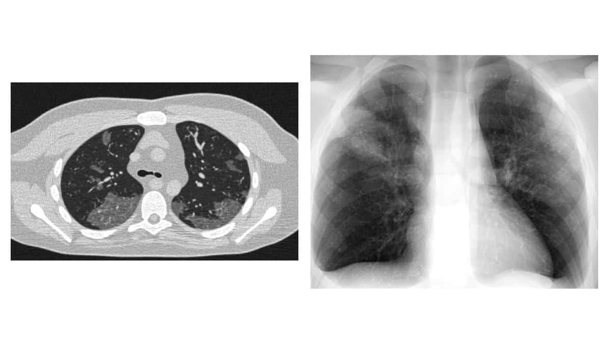
To model the specific abnormalities found in SARS-COV-2 patients, the group manually delineated the unique morphological features characteristic of COVID-19 in CT imaging data from 20 patients with confirmed infections. They then incorporated these features, known as ground-glass opacities, into the XCAT models. To make their phantom as realistic as possible, the researchers also modified the texture of each segmented abnormality to that of a material filled with fluid to match the observed pathologies.
… virtually imaged
The group produced three COVID-19 computational models with abnormalities of different shapes and locations within the lungs. The researchers then used these models together with a validated radiographic simulator (DukeSim) to obtain clinically realistic virtual CT and chest-radiography images. “Subjectively,” the authors explain, “the simulated abnormalities were realistic in terms of shape and texture.”

“We have developed strategies to adapt and use virtual imaging trials for imaging studies of COVID-19,” first author Ehsan Abadi concludes. “This will provide the foundation for the effective assessment and optimization of CT and radiography acquisitions and analysis tools to manage the COVID-19 pandemic.” Future work will focus on modelling different stages and manifestations of the disease, with the aim of optimizing screening and follow-up of patients.
