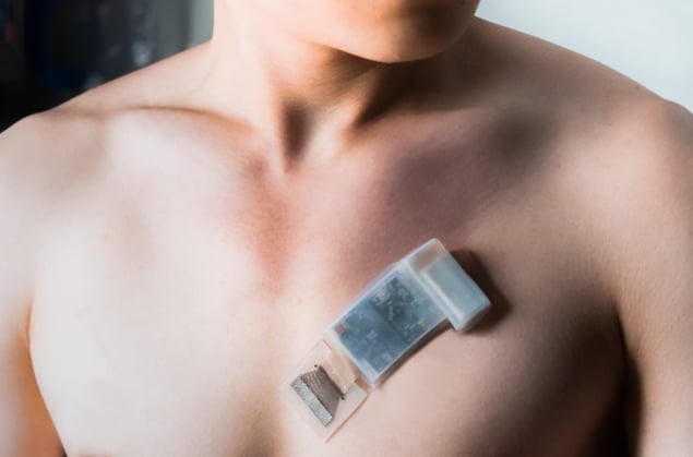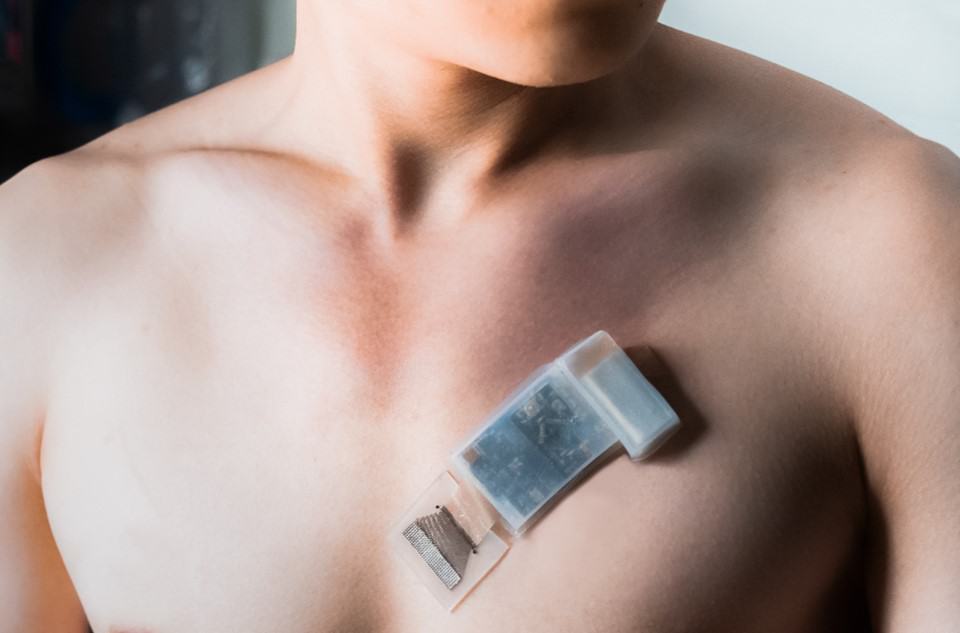
Researchers in the US have designed an ultrasound transducer that transmits information wirelessly and can be worn comfortably on the skin, overcoming two major shortcomings of previous devices. Developed by Muyang Lin, Sheng Xu and colleagues at the University of California San Diego (UCSD), the new transducer could be used to monitor patients with serious cardiovascular conditions, as well as to help athletes keep track of their training.
Ultrasound transducers work by transmitting high-frequency sound waves into the body, then detecting the waves reflected from tissues that have different densities and acoustic properties. Over the past several decades, improvements to probe and circuit designs, combined with better algorithms for processing ultrasound signals, have produced transducers that can conform to the folds of a person’s skin. This has allowed the devices to measure ultrasound signals continuously, which is especially useful for monitoring the pulsing of veins and arteries.
Researchers in Xu’s lab had previously developed wearable ultrasound probes that could monitor several physiological parameters of deep tissues, including blood pressure, blood flow and even cardiac imaging. Even so, the technology had some shortcomings. “These wearable probes are all wired to a bulky machine for power and data collection, and will shift in relative position during human motion, making them lose track of targets,” explains Lin, a PhD student in nanoengineering at UCSD and lead author of a paper in Nature Biotechnology on the device.
Because of these flaws, previous continuous ultrasound sensors could seriously inhibit a wearer’s mobility. They also required frequent readjustments as wearers moved around.
Ultrasound untethered
To address these problems, the UCSD team developed a new device based on a miniaturized, flexible control circuit that interfaces with an array of transducers. This device collects the ultrasound signals but does not process them directly. Instead, it relays them wirelessly to a computer or smartphone, which processes them using machine learning.
“We developed an algorithm to automatically analyse the signal and select the channel that has the best signal on moving target tissue,” Lin explains. “Therefore, the signals from the target tissue are continuous, even during human motion.”
The researchers tested this capability by using the device to track the position of a human subject’s carotid artery while monitoring the pulsation of blood within. This artery supplies blood to the head and neck, so they trained the algorithm to recognize displacements caused by different motions of the subject’s head.
Although the team only trained the algorithm on a single subject, a further advanced adaptation algorithm allowed new wearers to use the sensor with minimal retraining. Once trained, the device could detect ultrasound signals of the carotid artery’s pulsation as deep as 164 mm beneath the skin, even when the wearer was exercising.
Multi-use monitor
Xu and colleagues originally intended to test the sensor’s capabilities as a blood pressure monitor. Through their experiments, however, they discovered it could also monitor other important parameters, including arterial stiffness, the volume of blood pumped out by the heart and the amount of air exhaled by the wearer.

Wearable ultrasound sensor provides continuous cardiac imaging
Ultimately, the researchers predict their design could open up a wide range of possibilities for continuous ultrasound monitoring. “By using wearable ultrasound technology, we can untether the patient from bulky machines and automate the ultrasonic examinations,” Lin says. “Deep tissue physiology can be monitored in motion, which provides unprecedented opportunities for medical ultrasonography and exercise physiology.”
These capabilities could be life-changing for patients living with cardiovascular conditions, Lin says. “For at-risk populations, abnormal values of blood pressure and cardiac output at rest or during exercise are hallmarks of heart failure,” he explains. But the applications don’t end there. “For a healthy population, our device can measure cardiovascular responses to exercise in real-time. Thus, it can provide insights into the actual workout intensity exerted by each person, which can guide the formulation of personalized training plans.”
