
A new X-ray technique has revealed hitherto unknown structures in human bone. The technique, which uses a synchrotron beam to map the 3D orientation of nanocrystals and nanostructures within a material, advances our understanding of bone structure and how it relates to bone mechanics and bone disease. According to its developers, the method could also shed light on other natural and synthetic structures in which crystal orientation plays a role in determining the material’s properties.
Bone is an impressive natural material. It is both stiff, able to endure load without deforming much, and tough, meaning that it doesn’t break easily when bent or placed under load. Stiffness and toughness are rarely found together in a single material, and indeed bone gets its unusual properties from two main constituents: protein-based collagen fibrils, reinforced by nanocrystals of a calcium phosphate mineral called hydroxylapatite. These building blocks appear in various twisted patterns, and incorporate thin layers known as lamella that stack together like plywood. However, the precise way they fit together has proved difficult to understand because their organization is complex at every length scale.
Researchers from Aarhus University in Denmark, the European Synchrotron (ESRF) in France, Chalmers University in Sweden and the Paul Scherrer Institute in Switzerland have now used X-ray scattering data to show that the small-scale structure of bone is not uniform, as previously assumed, but contains deviations in the orientation of the hydroxylapatite nanocrystals with respect to the bone’s nanostructure. “To the best of our knowledge, this is the first time that that the ‘twisted plywood’ pattern of human lamellar bone has been mapped in 3D at such high resolution,” says Marianne Liebi, a materials physicist at Chalmers and the study’s co-first author.
Tensor tomography
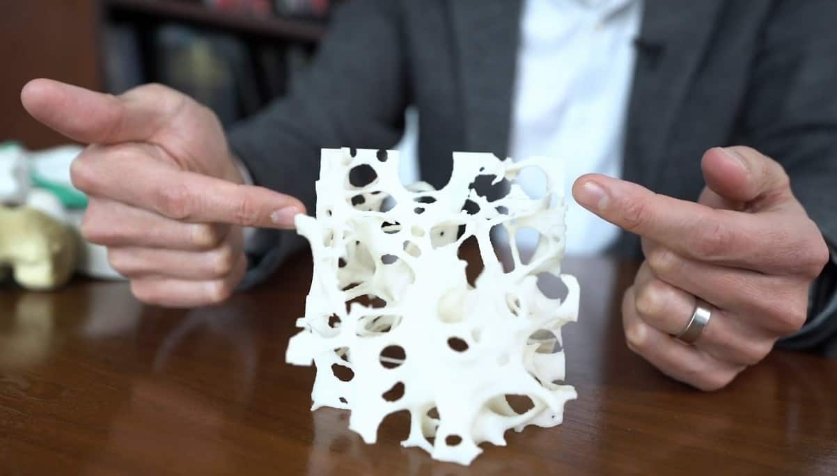
The researchers focused on a type of human bone known as trabecular bone, which is porous and found at the ends of long bones like the femur. The pores in the bone are connected by thin rods and plates of bone tissue.
In their experiments, members of the team scanned a 40x40x40 micron cross-section of trabecular bone across a beam of synchrotron radiation while rotating the sample around its x and y axes. By combining small-angle X-ray scattering and wide-angle X-ray scattering, they were able to probe the bone’s nanoscale structures and the atomic structure of its biomineral crystal lattice at the same time.
To extract information on the bone’s 3D structure from their x-y scan, they used a technique called tensor tomography to analyse the intensity distribution of the scattered radiation in reciprocal space (an imaginary space in which planes of atoms are represented by reciprocal points and all lengths are the inverse of their length in real space). If, for example, the crystals in the material are not randomly oriented, the intensity of the X-ray scattering signals will be large for some orientations and small for others, explains co-team leader Henrik Birkedal, who is in the Department of Chemistry and iNANO at Aarhus. “This can be likened to the presence of landmasses on a globe,” he says. “By mapping the variation of the scattering signals with sample orientation (corresponding to making a topographic map in the globe analogy), we can determine how the crystals or nanostructures are oriented in 3D.”
The scattered intensity “projection” of the x-y scan revealed a clear lamellar structure of oriented material, interlaced with disordered regions. According to Tilman Grunewald, the study’s other first author and a former ESRF scientist, “it’s a bit too early” to explain why the orientation of the hydroxylapatite nanocrystals is sometimes disordered, but the data nevertheless provides a focus for later research. “Essentially, our approach provides a new way of looking into the underlying structure of bone,” he says.
Synchrotron advances
Manfred Burghammer, the study’s co-leader and the scientist in charge of the ESRF’s ID13 beamline where the measurements were made, says that this new approach was made possible by “drastic progress” that is currently being made in X-ray synchrotrons. “We are expecting that the current upgrades of synchrotron sources will increase our power immensely in the next years,” he adds.
Breaking the mystery of bone micro-architecture
The team now plan to use their method to analyse bone material from patients with diseases such as osteoporosis and other disorders associated with age-related changes in bone structure, which can impair the bone’s mechanical properties. They also hope to study types of bone microstructure other than the one analysed in this work.
“Finally, we wish to understand the origin of the localized differences in orientation between nanostructure and crystals that we have observed,” Birkedal tells Physics World. “This will likely require improving the resolution of our technique by using smaller X-ray beams.”
Full details of the technique are reported in Science Advances.